
National Cancer Institute Immunotherapy Validation study confirmed Imago’s fractal-geometry-based image post-processing to reveal both the progression and regression of breast, lung, and glioblastoma cancer growth.
“I’m a Living Breathing Fractal and So are You” Ben Weiss / TED Talk
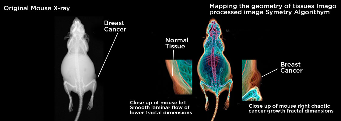
The analysis of three-dimensional imaging processes is very complicated. Normal human tissue has a different “texture” than cancer cell tissue due to their different geometric properties, and therefore will have a different fractal number. Different types of biological tissue will distinguish itself and have different curves than other tissue.
Transformation of original medical images into Imago’s fractal geometry patterns has been validated across many clinical imaging applications, involving different tissue types, imaging modalities, and disease states from breast to lung to vasculature.
- Geometric patterns were clearly distinguished and mathematically quantified between normal and cancerous lesions with X-ray, PET/CT, and MR images.
- Revealed the presence and extent of metastatic lung cancer using standard X- ray images as confirmed by pathology.
- Visualized and quantified changes in tumor sizes using geometric patterns and fractal dimensions consistent with immune responses.
- Validated diagnostic in vivo imaging for monitoring cancer tumor response to immunotherapy drug treatment in animals.