
On Average 6,000,000 dogs will be diagnosed with cancer this year.
The total number of companion animal vet clinics in the US alone is about 26,000 with a total estimated average of 100,000 diagnosis x-rays processed daily.
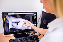
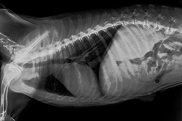
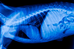
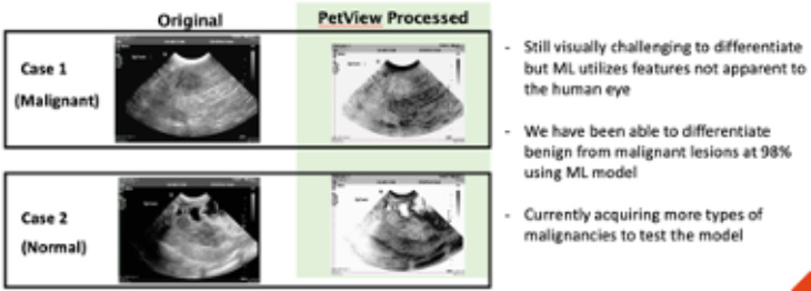
Veterinarians can use Imago’s branded PetView Reveal™ as an extension of their existing imaging tool set. Whether the clinic has X-ray systems, ultrasound, or both, the DICOM images are transferred directly to PetViewDX’s portal from a Veterinary’s digital system and processed in minutes. The returned images reveal margins and internal tissue structures not visible in the original image. Visual Intelligence software will categorize the image as normal/abnormal, and if abnormal, whether it is benign/malignant, with a confidence metric. By example, in the case of Splenic Mass Use Case, current results indicate separability of diagnosis between normal and abnormal spleens based on image processing followed by Machine Learning/Artificial Intelligence (ML/AI) scores with confidence level of 96% and malignant versus benign with a confidence level >75%.
Take Image
(Radiograph/ultrasound)
Upload via Web portal
Download processed image seconds later with Machine Learning Score when applicable
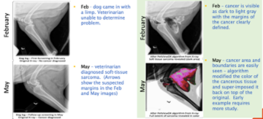
Soft Tissue Sarcoma
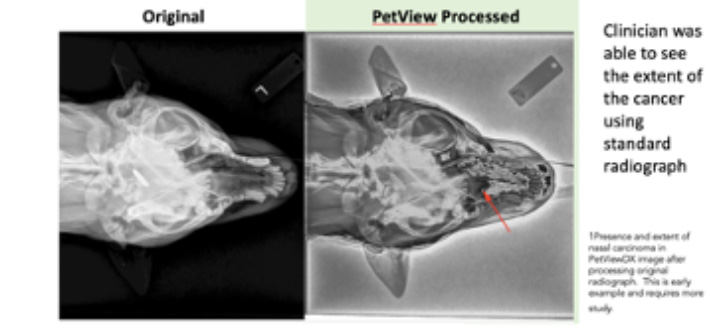
Nasal Carcinoma 1
Splenic Mass Differentiation
Imago is conducting a retrospective validation study with Auburn University utilizing 100 use cases in dogs to determine the accuracy of Visual Intelligence in measuring Splenic and Liver Mass differentiation. Imago projects an 85% normal/abnormal analysis meaning the features in the images uploaded correlated accurately or better with 85% of the machine learning models. The data from this validation study will support PetViewDX’s go-to-market strategy beginning 2022 targeting veterinary clinics, hospitals, universities, research institutions and emergency care centers.
Once this channel is developed and/or data with validation supports market opportunity, Imago anticipates spinning off this operating unit to a major strategic partner or entity servicing the global veterinary market.
For more information go to PetViewDX website; https://petviewdx.com